
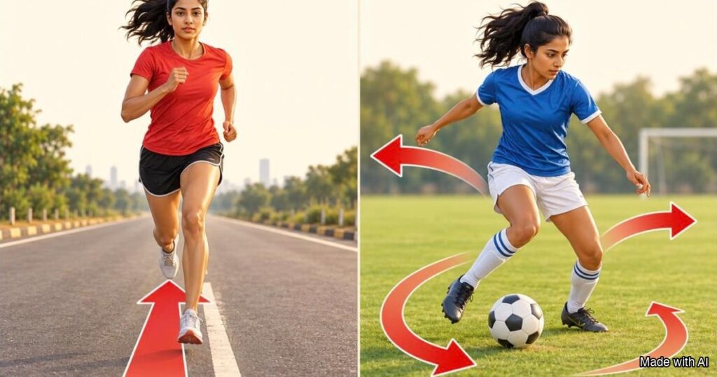
Short answer: Same bone, two sports. In a scan study of 32 college women, the shin bone of football players had 7.5% more area and was 29.4% stiffer in its weakest direction than the shin bone of long distance runners. The runners’ bone was longest front to back, along the push of running.
In this article
- Same bone, two sports: the shin bone of football players had 7.5% more area than runners’ in a scan study of 32 college women.
- It was 29.4% stiffer in its weakest direction, with an 8.2% higher estimated breaking load.
- Runners’ bone was longest front to back, along the push of running.
- A second study found 18.5 to 30% more bending resistance in 20 football players.
- Running builds bone too: a 30% higher strength index than in active women who did not train in a sport.
What the scan study compared
A scan study of 32 college women looked at one bone in two kinds of athletes: the shin bone, called the tibia. Sixteen of them were long distance runners and sixteen played football. The scans were taken in the middle of the bone shaft, and the researchers measured size, shape and estimated strength. They also corrected for the arm bone, so that people who were simply born with stronger bones would not skew the result.
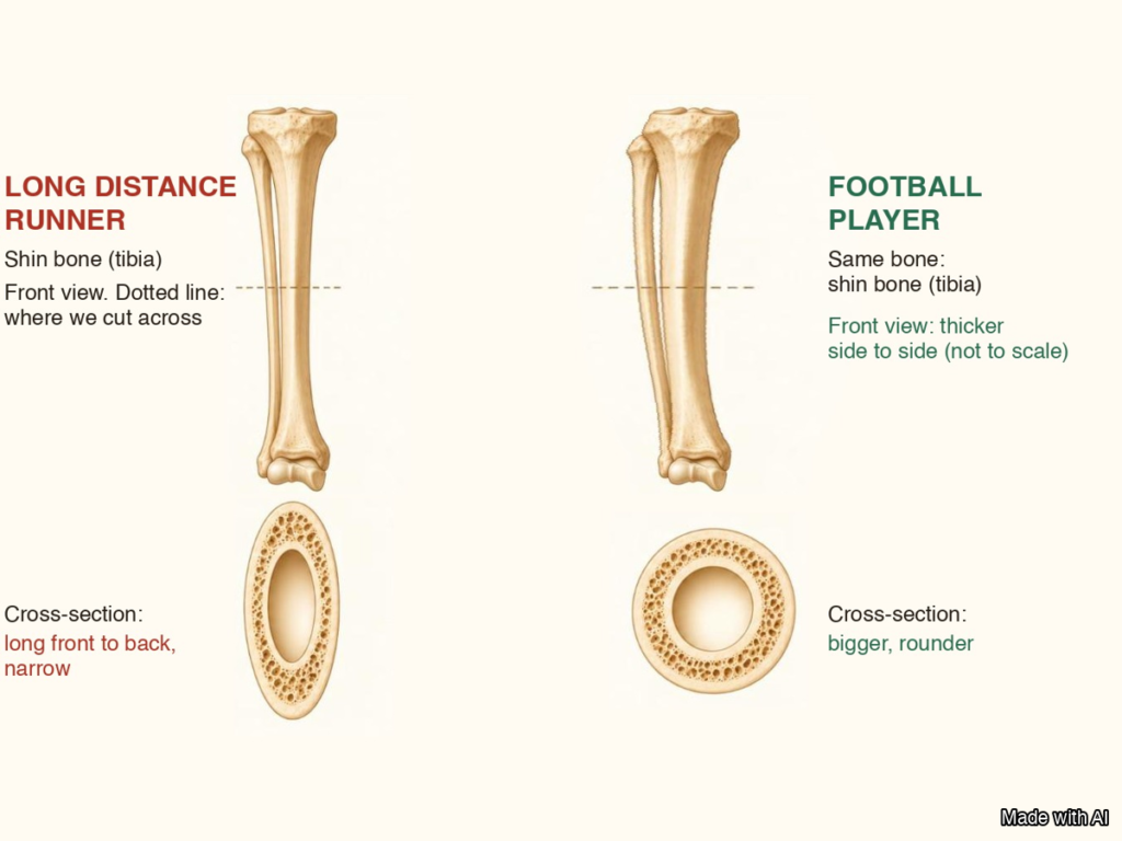
How the two shin bones look
The runners’ shin bones were close to normal reference values for size. Their shape was different, though. The cut-across shape was longer from front to back than side to side, so most of the bone material sat in the direction of the push. The authors link this to the one direction of loading in running. In their words, running seemed to cause only limited adaptation of the shin bone.
The football players’ shin bones were bigger. They had 7.5% more total bone area than the runners’ bones. They were 29.4% stiffer in their weakest direction. Their estimated breaking load was 8.2% higher. The cut-across shape stayed close to the normal reference shape, which the authors call a multiplane adaptation: the bone grew in many directions, not one.
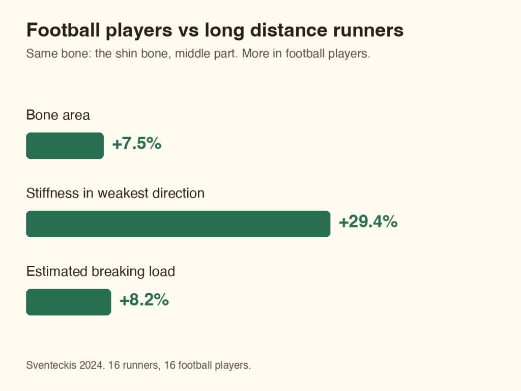
Why the direction of the push matters
Bone puts material where the load comes from. Running pushes the shin bone mostly in one direction, again and again. Football adds kicks, turns and side steps, so the push comes from more directions. The authors of the scan study link the different shapes to these different loads.
This is a link, not proof of cause. The study compared two groups at one point in time, in young women. It cannot say what each athlete’s bone looked like before they started their sport.
Three more findings
A second scan study compared 20 college women who play football with 20 matched women who did not. At three places along the shin bone, the football players had 18.5 to 30% more resistance to bending. Their estimated strength index was 16.5 to 19% higher. Their bone was a little less dense, by 1.5 to 3%, so the gain was in shape and size. The authors say trials are needed to confirm that football itself builds the bone.
Running is not bad for bones. In 221 women, the shin bone strength index was 30% higher in low-impact repeating sports such as running than in active women who did not train in a sport. For jumping sports it was 31% higher. For sports with odd impact, such as racket sports and football, it was 19% higher. Every kind of load beat no sport.
The other side of the story is what happens with no load. In a bed rest study, six healthy men rested in bed for 17 weeks. Their shin bone lost 2.2% of its mineral, and the heel bone lost 10.4%. Bone keeps what it is asked to carry.
What this means for you
No sport is wrong. Each one shapes the bone to its own load, and moving beats not moving. If you only do one kind of exercise, adding other kinds of movement changes the direction of the load.
With a heart problem, ask your doctor first. A physician supervised exercise intervention chooses the movements that are safe for you, and adds load step by step. Physician supervised, exercise based cardiac rehab works the same way: small, planned steps up. The runner’s side of this story is on the Telangana Runners blog: runner vs football shin bone.
Your questions, answered
How is a runner’s shin bone different from a football player’s?
In a scan study of 32 college women, the football players’ shin bone had 7.5% more area and was 29.4% stiffer in its weakest direction (Sventeckis 2024). The runners’ bone had more material from front to back. The study compared groups at one time, so it shows a link, not a cause.
Does running weaken the shin bone?
No. In 221 women, the shin bone strength index was 30% higher in running type sports than in active women who did not train in a sport (Rantalainen 2010). The study found that football and jumping sports built more, but running still built bone.
Why would football build a bigger shin bone?
The authors link it to the direction of the load. Running pushes the bone one way, while football adds kicks, turns and side steps (Sventeckis 2024). A second study agrees, but it says trials are needed to confirm that football itself is the cause (Hughes 2022).
What happens to bone without load?
Bone is lost. In a study of six healthy men who rested in bed for 17 weeks, the shin bone lost 2.2% of its mineral and the heel bone lost 10.4% (LeBlanc 1990). Bone keeps what it is asked to carry, so regular load, from any sport, helps keep it.
Build the load safely, with your doctor
A physician supervised exercise intervention sets the safe pace and adds load step by step. General advice: your own doctor knows your health.
info@cardiacrehab.com · www.CardiacRehab.com
Cardiac Rehab, 7B Journalist Colony, Film Nagar, Jubilee Hills, Hyderabad, Telangana 500096
References
- Sventeckis AM, Surowiec RK, Fuchs RK, Warden SJ. Cross-sectional size, shape, and estimated strength of the tibia, fibula and second metatarsal in female collegiate cross-country runners and football players. Bone. 2024;188:117233. pubmed.ncbi.nlm.nih.gov/39127437
- Hughes JM, Dickin DC, Wang H. Football participation is associated with benefits in tibial bone cross-sectional geometry and strength in young women. J Sports Med Phys Fitness. 2022;62(7):969-973. pubmed.ncbi.nlm.nih.gov/34498820
- Rantalainen T, Nikander R, Heinonen A, Suominen H, Sievanen H. Direction-specific diaphyseal geometry and mineral mass distribution of tibia and fibula: a pQCT study of female athletes representing different exercise loading types. Calcif Tissue Int. 2010;86(6):447-454. pubmed.ncbi.nlm.nih.gov/20383493
- LeBlanc AD, Schneider VS, Evans HJ, Engelbretson DA, Krebs JM. Bone mineral loss and recovery after 17 weeks of bed rest. J Bone Miner Res. 1990;5(8):843-850. pubmed.ncbi.nlm.nih.gov/2239368